XI. T Wave
Abnormalities
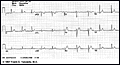
INTRODUCTION:The T wave is the most labile wave in the ECG. T wave changes
including low-amplitude T waves and abnormally inverted T waves
may be the result of many cardiac and non-cardiac conditions. The normal T wave
is usually in the same direction as the QRS except in the right precordial leads
(see V2 below). Also, the normal T wave is asymmetric with the first half
moving more slowly than the second half. In the normal ECG (see below) the T
wave is always upright in leads I, II, V3-6, and always inverted in lead aVR.
The other leads are variable depending on the direction of the QRS and the age
of the patient.
click here
to view
Differential Diagnosis of T Wave Inversion
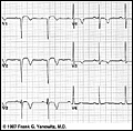
 Q wave and non-Q wave MI (e.g., evolving anteroseptal MI):
click
here to view
Myocardial ischemia
Subacute or old pericarditis
Myocarditis
Myocardial contusion (from trauma)
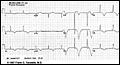
CNS
disease causing long QT interval (especially subarrachnoid hemorrhage; see
below):
click
here to view
Idiopathic apical hypertrophy (a rare form of hypertrophic
cardiomyopathy)
Mitral
valve prolapse
Digoxin effect
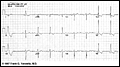
RVH
and LVH with "strain" (see below: T wave inversion in leads aVL, V4-6 in LVH)
click
here to view

)
)
)
)